
OCT Scanning in Wallsend
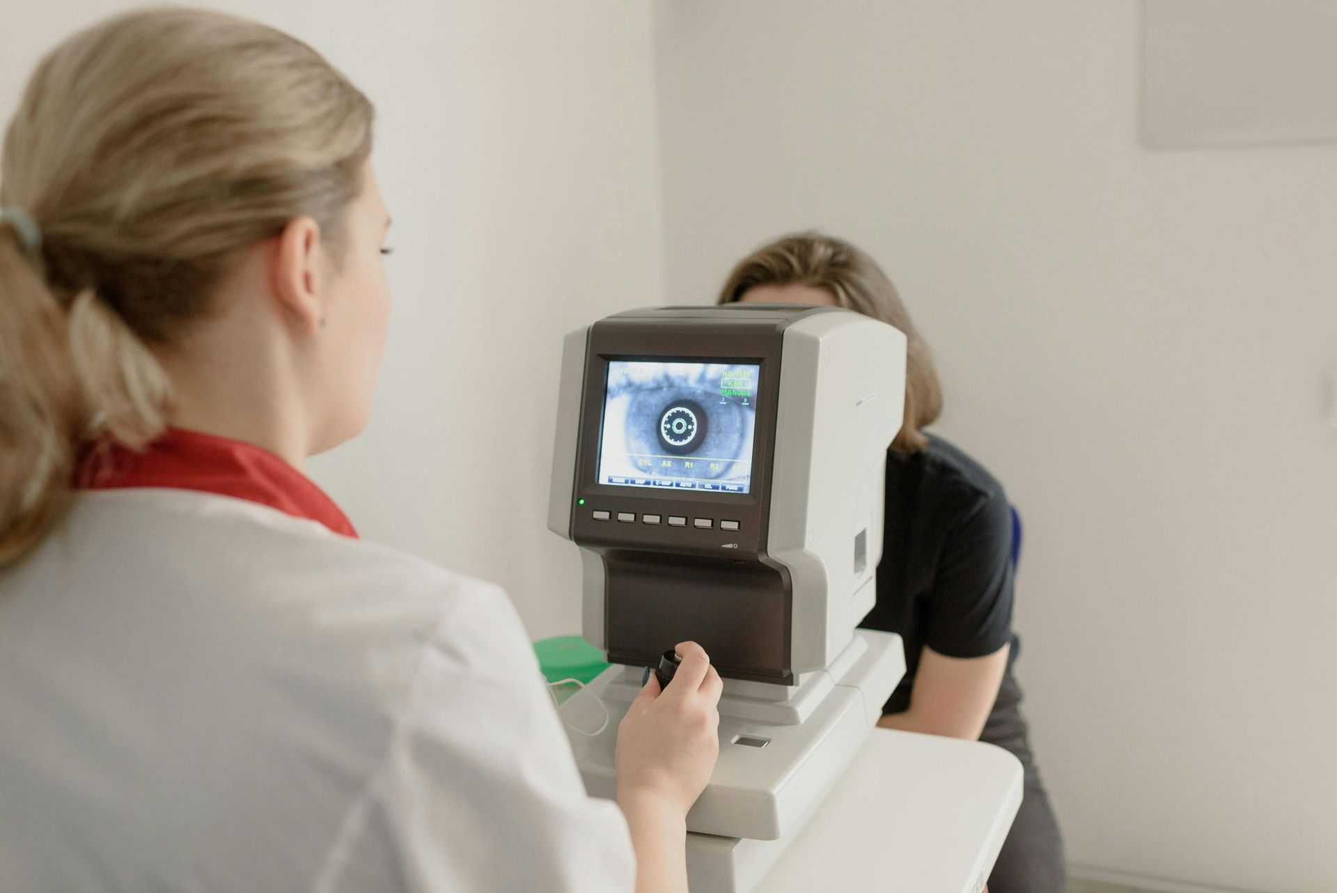
High-Resolution Retinal Imaging
OCT scanning allows us to examine the internal layers of the retina with exceptional detail, offering insight into structures that cannot be viewed through standard testing alone.
At EyeMax EyeCare in Wallsend we use OCT technology to capture cross-sectional images that help identify early changes in the optic nerve, macula and surrounding tissues. This type of imaging is valuable for monitoring conditions that may affect retinal thickness or layer integrity.
Our scans are performed using industry-recognised equipment designed to produce consistent, high-resolution results. The process is non-invasive, fast and suitable for patients who require ongoing monitoring over time. We guide you through every step so you understand what is being imaged and how the scan assists your ongoing eye care.
To book an OCT scan or learn more about the process, call
(02) 4955 9950.
These scans provide detailed retinal information that supports accurate review and long-term documentation of structural changes.
Book an Appointment
Thank you for contacting EyeMax EyeCare.
We will be in touch soon.
Oops, there was an error sending your message.
Please try again later.
Frequently Asked Questions
What does an OCT scan show?
An OCT scan provides cross-sectional images of the retina, showing the thickness, contour and structure of its layers in fine detail. The scan can reveal information about the macula, optic nerve and surrounding retinal tissue. It helps identify changes such as swelling, thinning or fluid accumulation. Because the scan displays each layer separately, it can highlight patterns not visible in a standard eye examination. These images allow clinicians to compare results with previous scans to track changes over time. OCT is commonly used to monitor conditions that influence retinal structure.
Is an OCT scan painful or uncomfortable?
An OCT scan is non-invasive and does not cause discomfort because it uses light to capture images rather than direct contact. During the scan you sit still and focus on a target while the device records images within a few seconds. There is no need for drops in most cases although dilation may sometimes be recommended to improve visibility. The process is suitable for people of various ages and requires minimal preparation. Because the scan is quick and gentle, many patients find it easy to complete during a routine appointment.
How long does an OCT scan take?
An OCT scan typically takes only a few minutes once you are positioned correctly at the device. The majority of the appointment involves ensuring that alignment is accurate so the captured images are clear. The scanning process itself is fast because the technology records multiple cross-sectional views in seconds. Additional time may be used to review the images and discuss findings. OCT is efficient enough to be completed during a standard eye examination, making it convenient for both initial assessment and ongoing monitoring.
How OCT Scanning Works
OCT scanning operates using light-based imaging technology that measures the depth and contour of retinal layers.
During the scan you look into the device while cross-sectional images are recorded without any contact with the eye. These images show the thickness and shape of layers such as the nerve fibre layer, photoreceptor regions and macular tissue. This level of detail helps identify subtle changes that may not be visible in traditional examinations. We review the scan together so you can see the structural information clearly and understand how it contributes to your assessment.
OCT is often used for monitoring conditions such as macula degeneration, glaucoma and diabetic eye changes, as each of these can affect retinal layers in specific ways. Results are stored securely so they can be compared at future appointments and used to track changes over time. This structured approach ensures consistent, informative imaging for ongoing review.
What to expect:
- Non-invasive cross-sectional imaging
- Detailed retinal layer assessment
- Clear documentation for future comparison


