
Glaucoma Test in Wallsend
Glaucoma Monitoring & Analysis
Glaucoma tests play a crucial role in identifying changes within the optic nerve, and at EyeMax EyeCare in Wallsend we carry out detailed assessments using recognised diagnostic tools.
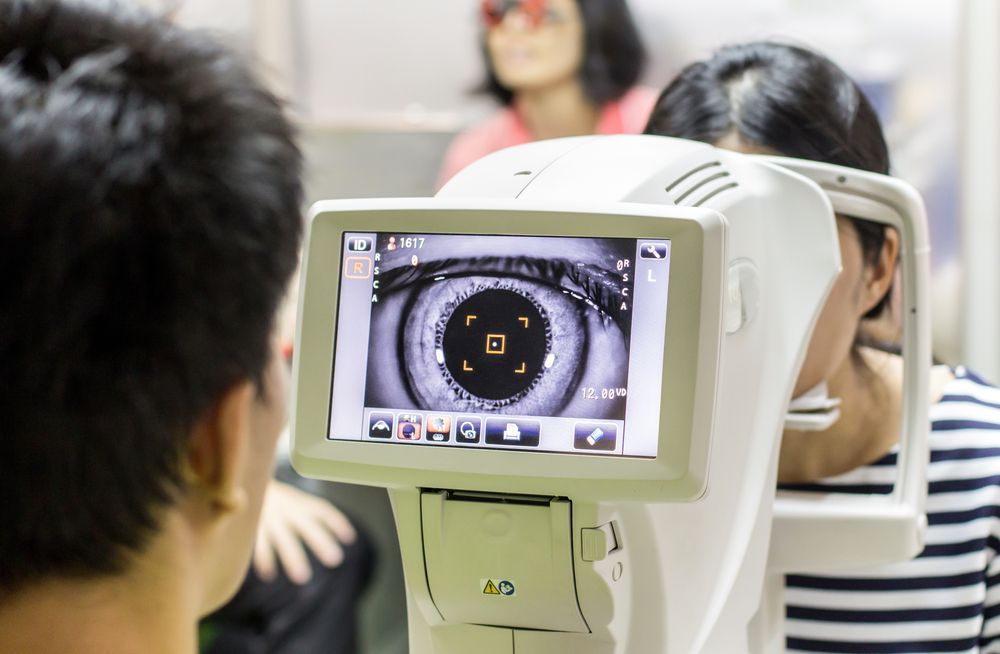
Our testing approach combines pressure readings, optic nerve imaging and visual field analysis to form a clear picture of how the nerve is functioning. We use tonometry to measure intraocular pressure, OCT to capture high-resolution scans of the optic nerve head and peripheral vision testing to detect pattern changes that may require monitoring. These tools allow us to evaluate structural and functional data together, providing a detailed understanding of how the eye is performing over time.
We explain each step clearly so you understand what is being measured and why these results matter for long-term eye health.
To make an appointment or ask questions about glaucoma testing, call
(02) 4955 9950.
Regular screening provides essential information that supports consistent monitoring and accurate comparison during future visits.
Book an Appointment
Thank you for contacting EyeMax EyeCare.
We will be in touch soon.
Oops, there was an error sending your message.
Please try again later.
Frequently Asked Questions
What is involved in a glaucoma test?
A glaucoma test usually includes several components that assess both the structure and function of the optic nerve. Tonometry measures the pressure inside the eye, OCT imaging captures detailed views of nerve fibres and visual field testing evaluates peripheral vision. These assessments work together to provide an understanding of how the optic nerve is functioning. Additional examinations may include slit lamp review of the drainage angle or retinal structure. Each test provides different information that helps document changes over time and supports long-term monitoring.
Can glaucoma develop without symptoms?
Glaucoma often develops without noticeable symptoms in its early stages, which is why routine testing is important. The condition typically affects peripheral vision first, and gradual changes can go unnoticed because the brain adapts to filling in gaps. Structural changes in the optic nerve may also progress quietly before any functional vision loss occurs. Diagnostic tools such as OCT imaging and visual field testing allow these changes to be detected early. Because symptoms may not appear until the condition advances, regular testing supports tracking subtle patterns over time.
What tools are most commonly used in glaucoma assessments?
Common tools used in glaucoma testing include tonometers for measuring intraocular pressure, OCT devices for imaging optic nerve structure and visual field machines for evaluating peripheral awareness. Slit lamp examinations help assess the drainage angle, while retinal photography captures structural detail for comparison. Each instrument contributes a different layer of information. OCT scans offer detailed structural insights, while visual field testing examines how the optic nerve performs functionally. This combination of tools helps create a comprehensive assessment that supports long-term monitoring.
How We Assess Glaucoma Risk
Our glaucoma testing process includes several clinically recognised evaluations that together form a comprehensive assessment.
We begin with a review of vision clarity and eye pressure measurements, which help establish a baseline for comparison. OCT imaging is used to capture detailed cross-sectional views of the optic nerve and surrounding retinal layers. These scans allow us to identify nerve fibre thickness and structural patterns that may require observation. Visual field testing is then completed to assess peripheral awareness, which can highlight functional changes that develop gradually.
We also examine the drainage angle where fluid moves through the eye because this can influence eye pressure dynamics. Throughout the assessment we provide clear explanations so you understand how each test contributes to the overall analysis. This structured approach ensures that findings are documented consistently for long-term monitoring.
Service highlights:
- Detailed optic nerve imaging
- Structured pressure and field testing
- Consistent long-term documentation


